
The vital crosstalk between breath and brain

If you’re lucky enough to live to 80, you’ll take up to a billion breaths in the course of your life, inhaling and exhaling enough air to fill about 50 Goodyear blimps or more. We take about 20,000 breaths a day, sucking in oxygen to fuel our cells and tissues and ridding the body of carbon dioxide that builds up as a result of cellular metabolism. Breathing is so essential to life that people generally die within minutes if it stops.
It’s a behavior so automatic that we tend to take it for granted. But breathing is a physiological marvel — both extremely reliable and incredibly flexible. Our breathing rate can change almost instantaneously in response to stress or arousal and even before an increase in physical activity. And breathing is so seamlessly coordinated with other behaviors like eating, talking, laughing and sighing that you may never have even noticed how your breathing changes to accommodate them. Breathing also can influence your state of mind, as evidenced by the controlled breathing practices of yoga and other ancient meditative traditions.
In recent years, researchers have begun to unravel some of the underlying neural mechanisms of breathing and its many influences on body and mind. In the late 1980s, neuroscientists identified a network of neurons in the brainstem that sets the rhythm for respiration. That discovery has been a springboard for investigations into how the brain integrates breathing with other behaviors. At the same time, researchers have been finding evidence that breathing may influence activity across wide swaths of the brain, including ones with important roles in emotion and cognition.
“Breathing has a lot of jobs,” says Jack L. Feldman, a neuroscientist at the University of California, Los Angeles, and co-author of a recent article on the interplay of breathing and emotion in the Annual Review of Neuroscience. “It’s very complicated because we’re constantly changing our posture and our metabolism, and it has to be coordinated with all these other behaviors.”
Each breath a symphony of lung, muscle, brain
Every time you inhale, your lungs fill with oxygen-rich air that then diffuses into your bloodstream to be distributed throughout your body. A typical pair of human lungs contains about 500 million tiny sacs called alveoli, the walls of which are where gases pass between the airway and bloodstream. The total surface area of this interface is about 750 square feet — a bit more than the square footage of a typical one-bedroom apartment in San Francisco and a bit less than that of a racquetball court.
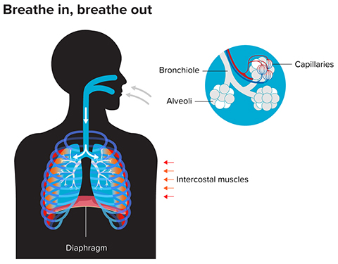
“The remarkable thing about mammals, including humans, is that we pack an enormous amount of surface area into our chests,” says Feldman. More surface area means more gas is exchanged per second.
But the lungs can’t do it alone. They’re essentially limp sacks of tissue. “In order for this to work, the lungs have to be pumped like a bellows,” Feldman says. And they are — with each inhalation, the diaphragm muscle at the bottom of the chest cavity contracts, moving downward about half an inch. At the same time, the intercostal muscles between the ribs move the rib cage up and out — all of which expands the lungs and draws in air. (If you’ve ever had the wind knocked out of you by a blow to the stomach, you know all about the diaphragm; and if you’ve eaten barbecued ribs, you have encountered intercostal muscles.)
At rest, these muscles contract only during inhalation. Exhalation occurs passively when the muscles relax and the lungs deflate. During exercise, different sets of muscles contract to force out air and speed up respiration.
Unlike the heart muscle, which has pacemaker cells that set its rhythm, the muscles that control breathing take their orders from the brain. Given the life-enabling importance of those brain signals, it took a surprisingly long time to track them down. One of the first to ponder their source was Galen, the Greek physician who noticed that gladiators whose necks were broken above a certain level were unable to breathe normally. Later experiments pointed to the brainstem, and in the 1930s, the British physiologist Edgar Adrian demonstrated that the dissected brainstem of a goldfish continues to produce rhythmic electrical activity, which he believed to be the pattern-generating signal underlying respiration.
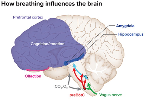
But the exact location of the brainstem respiratory-pattern generator remained unknown until the late 1980s, when Feldman and colleagues narrowed it down to a network of about 3,000 neurons in the rodent brainstem (in humans it contains about 10,000 neurons). It’s now called the pre-Bötzinger complex, or preBötC. Neurons there spontaneously exhibit rhythmic bursts of electrical activity that, relayed through intermediate neurons, direct the muscles that control breathing.
Over the years, some people have assumed Bötzinger must have been a famous anatomist, Feldman says, perhaps a German or Austrian. But in fact the name came to him in a flash during a dinner at a scientific conference where he suspected a colleague was inappropriately about to claim the discovery for himself. Feldman clinked his glass to propose a toast and suggested naming the brain region after the wine being served, which came from the area around Bötzingen, Germany. Perhaps lubricated by said wine, the others agreed, and the name stuck. “Scientists are just as weird as anyone else,” Feldman says. “We have fun doing things like this.”
Pinpointing breath’s rhythm setters
Much of Feldman’s subsequent research has focused on understanding exactly how neurons in the preBötC generate the breathing rhythm. This work also has laid a foundation for his lab and others to investigate how the brain orchestrates the interplay between breathing and other behaviors that require alterations in breathing.
Sighing is one interesting example. A long, deep breath can express many things: sadness, relief, resignation, yearning, exhaustion. But we humans aren’t the only ones who sigh — it’s thought that all mammals do — and it may be because sighing has an important biological function in addition to its expressive qualities. Humans sigh every few minutes, and each sigh begins with an inhale that takes in about twice as much air as a normal breath. Scientists suspect this helps pop open collapsed alveoli, the tiny chambers in the lung where gas exchange occurs, much as blowing into a latex glove pops open the fingers. Several lines of evidence support this idea: Hospital ventilators programmed to incorporate periodic sighing, for example, have been shown to improve lung function and maintain patients’ blood oxygen levels.
In a study published in 2016 in Nature, Feldman and colleagues identified four small populations of neurons that appear to be responsible for generating sighs in rodents. Two of these groups of neurons reside in a brainstem region near the preBötC, and they send signals to the other two groups, which reside inside the preBötC. When the researchers killed these preBötC neurons with a highly selective toxin, the rats ceased to sigh, but their breathing remained robust. On the other hand, when scientists injected neuropeptides that activate the neurons, the rats sighed 10 times more frequently. In essence, the researchers concluded, these four groups of neurons form a circuit that tells preBötC to interrupt its regular program of normal-sized breaths and order up a deeper breath.
The preBötC also has a role in coordinating other behaviors with breathing. One of Feldman’s collaborators on the sighing paper, neuroscientist Kevin Yackle, and colleagues recently used mice to investigate interactions between breathing and vocalizations. When separated from their nest, newborn mice make ultrasonic cries too high-pitched for humans to hear. There are typically several cries at regular intervals within a single breath, not unlike the syllables in human speech, says Yackle, who’s now at the University of California, San Francisco. “You have this slower breathing rhythm and then nested within it you have this faster vocalization rhythm,” he says.
To figure out how this works, the researchers worked their way backward from the larynx, the part of the throat involved in producing sound. They used anatomical tracers to identify the neurons that control the larynx and follow their connections back to a cluster of cells in the brainstem in an area they named the intermediate reticular oscillator, or iRO. Using a variety of techniques, the researchers found that killing or inhibiting iRO neurons removes the ability to vocalize a cry, and stimulating them increases the number of cries per breath.
When the researchers dissected out slices of brain tissue with iRO neurons, the cells kept firing in a regular pattern. “These neurons produce a rhythm that’s exactly like the cries in the animal, where it’s faster than but nested within the preBötC breathing rhythm,” Yackle says.
Additional experiments suggested that iRO neurons help integrate vocalizations with breathing by telling the preBötC to make tiny inhalations that interrupt exhalation — enabling a series of brief cries to fit neatly within a single exhaled breath. That is, rhythmic crying isn’t produced by a series of exhalations but rather by one long exhalation with several interruptions.
The findings, reported earlier this year in Neuron, may have implications for understanding human language. The number of syllables per second falls within a relatively narrow range across all human languages, Yackle says. Perhaps, he suggests, that’s due to constraints imposed by the need to coordinate vocalizations with breathing.
Setting the pace in the brain
Recent studies have suggested that breathing can influence people’s performance on a surprisingly wide range of lab tests. Where someone is in the cycle of inhalation and exhalation can influence abilities as diverse as detecting a faint touch and distinguishing three-dimensional objects. One study found that people tend to inhale just before a cognitive task — and that doing so tends to improve performance. Several have found that it is only breathing through the nose that has these effects; breathing through the mouth does not.
One emerging idea about how this might work focuses on well-documented rhythmic oscillations of electrical activity in the brain. These waves, often measured with electrodes on the scalp, capture the cumulative activity of thousands of neurons, and for decades some neuroscientists have argued that they reflect communication between far-flung brain regions that could underlie important aspects of cognition. They could be, for example, how the brain integrates sensory information processed separately in auditory and visual parts of the brain to produce what we experience as a seamless perception of a scene’s sounds and sights. Some scientists even have proposed that such synchronized activity could be the basis of consciousness itself (needless to say, this has been hard to prove).
Growing evidence suggests breathing may set the pace for some of these oscillations. In experiments with rodents, several research teams have found that the breathing rhythm influences waves of activity in the hippocampus, a region critical for learning and memory. During wakefulness, the collective electrical activity of neurons in the hippocampus rises and falls at a consistent rate — typically between six and 10 times per second. This theta rhythm, as it’s called, occurs in all animals that have been studied, including humans.
In a 2016 study, neuroscientist Adriano Tort at the Federal University of Rio Grande do Norte in Brazil and colleagues set out to study theta oscillations but noticed that their electrodes also were picking up another rhythm, a slower one with about three peaks per second, roughly the same as a resting mouse’s respiration rate. At first they worried it was an artifact, Tort says, perhaps caused by an unstable electrode or the animal’s movements. But additional experiments convinced them that not only was the rhythmic activity real and synched with respiration, but also that it acted like a metronome to set the pace for the faster theta oscillations in the hippocampus.
Around the same time, neuroscientist Christina Zelano and colleagues reported similar findings in humans. Using data from electrodes placed by surgeons on the brains of epilepsy patients to monitor their seizures, the researchers found that natural breathing synchronizes oscillations within several brain regions, including the hippocampus and the amygdala, an important player in emotional processing. This synchronizing effect diminished when the researchers asked subjects to breathe through their mouths, suggesting that sensory feedback from nasal airflow plays a key role.
Not only does the respiration rhythm synchronize activity in brain regions involved in emotion and memory, it can also affect people’s performance on tasks involving emotion and memory, Zelano and colleagues found. In one experiment, they monitored subjects’ respiration and asked them to identify the emotion expressed by people in a set of photos developed by psychologists to test emotion recognition. Subjects were quicker to identify fearful faces when the photo appeared as they were taking a breath compared to during exhalation. In a different test, subjects more accurately remembered whether they’d seen a photo previously when it was presented as they inhaled. Again, the effects were strongest when subjects breathed through the nose.

More recent work suggests the respiratory rhythm could synchronize activity not just within but also between brain regions. In one study, neuroscientists Nikolaos Karalis and Anton Sirota found that the respiration rate synchronizes activity between the hippocampus and the prefrontal cortex in sleeping mice. This synchronization could play a role in making long-term memories, Karalis and Sirota suggest in a paper published earlier this year in Nature Communications. Many neuroscientists think memories initially form in the hippocampus before being transferred during sleep to the cortex for long-term storage — a process thought to require synchronized activity between the hippocampus and cortex.
For Tort, such findings suggest there may be important links between respiration and brain function, but he says more work is needed to connect the dots. The evidence that breathing influences brain oscillations is strong, he says. The challenge now is figuring out what that means for behavior, cognition and emotion.
Controlled breath, calm mind?
For millennia, practitioners of yoga and other ancient meditation traditions have practiced controlled breathing as a means of influencing their state of mind. In recent years, researchers have become increasingly interested in the biological mechanisms of these effects and how they might be applied to help people with anxiety and mood disorders.
One challenge has been separating the effects of breathing from other aspects of these practices, says Helen Lavretsky, a psychiatrist at UCLA. “It’s really hard to distinguish what’s most effective when you’re doing this multicomponent intervention where there’s stretching and movement and visualization and chanting,” she says. Not to mention the cultural and spiritual components many people attach to the practice.
For many years, Lavretsky has collaborated with neuroscientists and others to investigate how different types of meditation affect the brain and biological markers of stress and immune function. She has found, among other things, that meditation can improve performance on lab tests of memory and alter brain connectivity in older people with mild cognitive impairment, a potential precursor to Alzheimer’s disease and other types of dementia. In more recent studies, which have yet to be published, she’s moved toward investigating whether the breath control methods alone can help.
“Even though I’m a psychiatrist, my research is on how to avoid (prescribing) drugs,” says Lavretsky, who is also a certified yoga instructor. She thinks breathing exercises might be a good alternative for many people, especially with more research on which breathing techniques work best for which conditions and how they might be tailored to individuals. “We all have this tool, we just have to learn how to use it,” she says.
This article is republished from knowablemagazine.org.
Enjoy reading ASBMB Today?
Become a member to receive the print edition four times a year and the digital edition monthly.
Learn moreGet the latest from ASBMB Today
Enter your email address, and we’ll send you a weekly email with recent articles, interviews and more.
Latest in Science
Science highlights or most popular articles

Mitochondria shape kidney cell function
Researchers at the University of Washington, Seattle present the first quantitative comparison of mitochondrial interactomes between two epithelial cell types in the kidney.

Long-chain polyunsaturated fatty acids linked to postoperative delirium risk
Researchers show that altered lipid metabolism may contribute to postoperative delirium, a condition linked to increased risk for long-term cognitive decline. The study explores potential disease mechanisms, which have yet to be understood.

Glycosylation patterns across antibody isotypes distinguish tuberculosis states
Researchers at Taipei Medical University present the first site-specific glycosylation analysis of immunoglobulins in elderly tuberculosis patients.

Blood glycome possibly predicts lifespan
Researchers at the University of Santiago de Compostela show that total serum N-glycome can predict mortality independent of traditional risk factors.

Building a better model for drug delivery across the blood–brain barrier
Industry and academic scientists collaborated to develop a rat with humanized iron-transport receptors, enabling research into iron homeostasis and drugs that cross the brain’s barrier.

Fat synthesis enzyme crucial for milk fat and newborn growth
Researchers found that a deficiency of the fatty acid synthesis enzyme stearoyl-CoA desaturase-1 reduced mammary gland function during lactation and caused low birth weight in newborns that were fed milk from enzyme-deficient glands.