Avoiding common figure errors in manuscript submissions

When a manuscript is provisionally accepted at the Journal of Biological Chemistry, the figures are reviewed for quality and compliance with ASBMB editorial policies and JBC guidelines. If issues arise, the data integrity team contacts authors to request original files, revised figures or clarification about image features.
This review helps prevent inaccuracies from entering the scientific record and reduces the need for research‑integrity interventions later.
The volume of issues is higher than many authors expect.
In 2025, about one in 20 submitted manuscripts contained figure issues that would have required a correction had they been discovered after publication. The data integrity team also contacted authors of one‑third of all accepted manuscripts to address general presentation issues.
On average, resolving these requests added nearly a week to the time to final acceptance.
The process is overwhelmingly successful. Only a small number of manuscripts were declined due to serious concerns; in most cases, authors, editors and the data integrity team worked together to bring figures into compliance.
Fortunately, most figure issues can be prevented. Authors can minimize delays by reviewing JBC guidelines early and assembling figures accordingly.
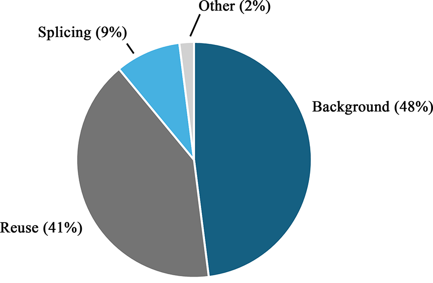
Here, we review the three most common infractions from the past year, background signal, image reuse and unreported splicing, and our recommended means for avoiding them.
Background signal errors
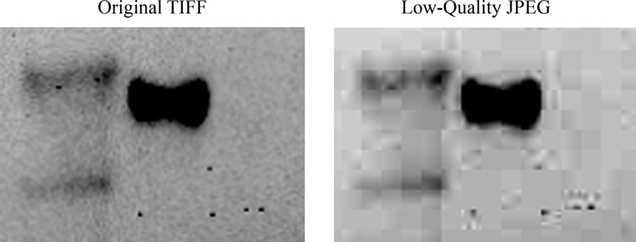
Background signal must be present in the original image capture and preserved throughout figure assembly. Saving files in lossless, high‑resolution formats such as TIFF, PDF or PNG is essential for preserving image quality.
Other image formats, such as JPEG, simplify images to reduce file size through “lossy” compression.
The JPEG algorithm prioritizes changes in brightness and strong edges — features usually absent from the background of typical gels, Western blots or fluorescence micrographs. As a result, large blocks of uniform pixel intensity can appear, creating the impression that the background may have been modified or spliced.
To maintain signal and resolution, the image must be exported from the imager in a lossless format, and every subsequent version must also be saved in a lossless format. If original TIFFs are arranged in an editing software and then exported as a JPEG, quality will still be lost.
Absent or insufficient background signal accounted for almost half of all queries to JBC authors. Image background is important for:
- Providing context for signal intensity and the quality of the labeling. If the contrast of a blot has been adjusted to the extent that the image consists of solid black bands on a solid white background, this information has been lost.
- Identifying other issues. Raw data may be requested to ensure that the lack of background signal is not obscuring other adjustments.
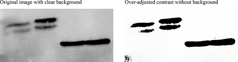
We can think of a Western blot as conceptually similar to a 96‑well plate. The difference is that the wells are pixels and the pixel intensities range from 0 (black) to 255 (white) for an 8-bit grayscale image. When contrast is adjusted, values closer to 0 are driven down to 0 and values closer to 255 are driven up to 255. While this is often done for Western blots, we would (hopefully) feel uncomfortable making a similar polarizing adjustment to absorbance values in a 96-well plate.
One argument is that what matters is simply whether the protein is present, but if this were the case, we could instead present Western blot data as a table with a row of lane numbers across the top and “positive” or “negative” across the rows for each protein. This table, of course, would provide no information about subtle shifts in size, relative abundance, or nonspecific labeling. The same is true for over-adjusted images.
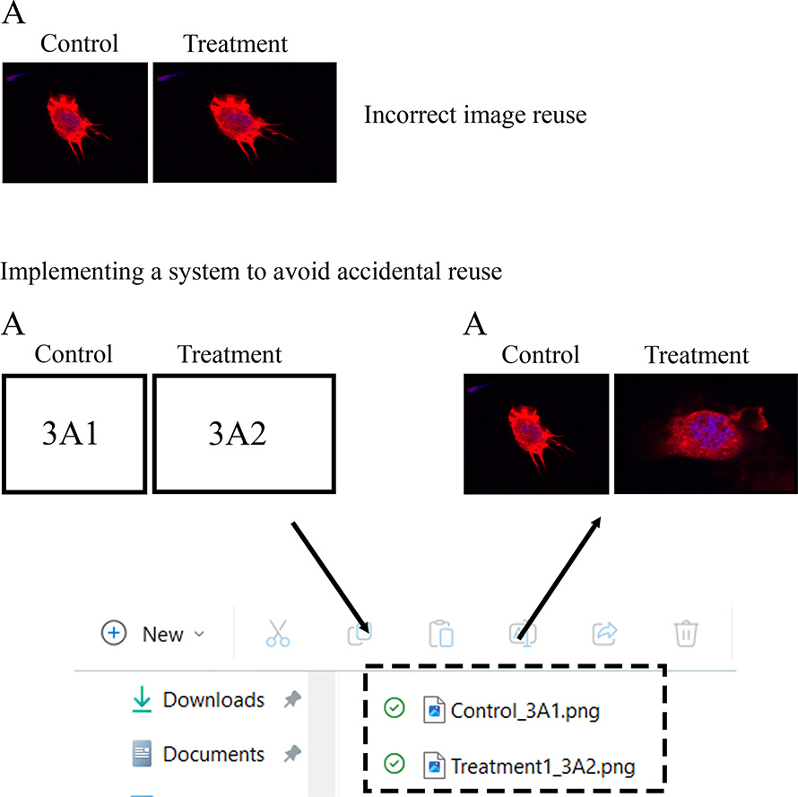
Image reuse errors
Nearly half of image‑reuse cases would have required a correction if identified after publication.
In most situations, images were reused accidentally or appeared in panels representing different experimental conditions. When authors could provide raw data to demonstrate the error, the figure was replaced with a corrected version.
The most common cause of these errors is file‑management issues. Some figures contain many similar panels, and it is easy to overlook a repeated image.
To prevent oversights like this, authors should use a reliable system for organizing image files. Each image should be assigned a unique, descriptive filename, and folders should be organized in a clear, consistent way.
Authors should avoid copying and pasting panels, as this increases the risk of accidental reuse and makes file‑tracking more difficult.
Image reuse can, of course, be useful for making data comparisons across figures. This type of reuse is acceptable, but ASBMB requires that it be stated explicitly in the figure legend. If the legend does not indicate the reuse, a revised manuscript will be requested.
As illustrated below, it may help to design figures in advance and assign each panel a serial number that appears in the image filename. If accidental reuse persists, authors may want to use image‑analysis software to screen figures before submission.
Undeclared splicing errors
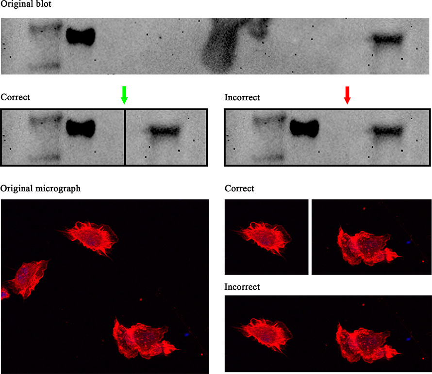
Undeclared splicing prompted about 10 percent of queries to authors in 2025.
While less common, splicing remains an important issue because it can cast doubt on a dataset.
If an image must be spliced — for example, to remove irrelevant lanes in a blot or gel — JBC guidelines require that the splice be clearly labeled and explained in the legend. This requirement also applies to micrographs, even when the individual images represent different channels, and splicing segments from separate image captures is generally not permitted.
Image reuse across different conditions is fundamentally incorrect because it alters the data itself, while an absent background signal or unlabeled splicing typically requires changes only to presentation, not the underlying results. Still, transparent image presentation, including visible background and clear labeling, communicates how data were collected and reinforces confidence in the findings. The JBC guidelines provide a framework to help authors present accurate, informative figures that uphold transparency and trust.
Enjoy reading ASBMB Today?
Become a member to receive the print edition four times a year and the digital edition monthly.
Learn moreGet the latest from ASBMB Today
Enter your email address, and we’ll send you a weekly email with recent articles, interviews and more.
Latest in Science
Science highlights or most popular articles

Fat synthesis enzyme crucial for milk fat and newborn growth
Researchers found that a deficiency of the fatty acid synthesis enzyme stearoyl-CoA desaturase-1 reduced mammary gland function during lactation and caused low birth weight in newborns that were fed milk from enzyme-deficient glands.

Flipping lipids and slime molds
A dull first job nearly pushed JBC associate editor Todd Graham out of science. Then a slime mold project changed his path. Now, he studies membrane biology and reflects on discovery, persistence and mentoring through uncertainty.

How smelling death alters worm behavior
Researchers have found that the roundworm C. elegans can smell death, and it changes how the worms behave, reproduce and age.

A chance encounter with the lab
Payton Stevens never planned to become a pancreatic cancer researcher. A temporary job set him on a path from rural Kentucky to leading research on Wnt signaling and metastasis, where he now pairs discovery with mentorship and science advocacy.

Light-activated small molecule could transform eye infection treatment
Contact lenses raise the risk of infectious keratitis, a leading cause of blindness worldwide. A biotech company is commercializing a light-activated therapy using a ROS-generating molecule to rapidly kill microbes in the cornea to preserve vision.
The molecular orchestra of memory
Calcium, calmodulin and calcium/calmodulin-dependent kinase II form a molecular axis that turns fleeting neural activity into lasting memories. New research shows how memories are stabilized, and possibly even protected or repaired.