Scientists use X-ray beams to determine role of zinc in development of ovarian follicles

To make a baby, first you need an egg. To have an egg, there needs to be a follicle. And in the very beginning of follicle development, there needs to be zinc.
The last of those statements represents the new findings reported recently by a team of researchers from Michigan State University, Northwestern University and the U.S. Department of Energy’s Argonne National Laboratory. The research builds upon earlier work looking at the role of zinc in fertilization and uncovers the importance of the metal earlier in the process of ovulation.
The results were reported in a paper in the Journal of Biological Chemistry that looked at the role of zinc in follicle development. The researchers, led by Teresa Woodruff and Tom O’Halloran of Michigan State University, used the Bionanoprobe at Argonne’s Advanced Photon Source to examine zinc and other trace elements in the egg cell itself as well as surrounding somatic cells. The APS is a DOE Office of Science user facility at Argonne.
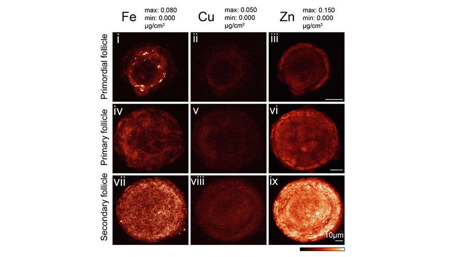
The researchers observed that the concentration of zinc in mouse follicles more than triples as they go through their development, while concentrations of other trace elements such as iron and copper increased to a lesser extent. The researchers also noticed that growing follicles acquired zinc at a rate 70 times faster than undeveloped follicles, suggesting a key role for zinc in the maturation of ovarian follicles.
“We had known from earlier research that zinc played an important role in fertilization, but this new research shows that it’s involved way before egg meets sperm,” said Argonne staff scientist Si Chen, an author of the study.
“This study is a beautiful example of how synchrotron-based science can open new fundamental understandings of biology, of reproduction, and of metabolism of disease,” said Michigan State microbiology professor Tom O’Halloran. “When you’re working with photons and X-ray beams, you might not always initially understand the connection to actual physiology — but this is really at the cutting edge of bringing new tools to the biology community.”
The Bionanoprobe is one of a few facilities in the world where researchers can measure the concentration of different elements within a cell’s organelles in cryogenic conditions, Chen said. “Being able to resolve subcellular compartments is important for understanding what’s going on behind the scenes as the follicle develops,” she said.
The importance of zinc for follicle development is related to the fact that there are zinc-activated proteins that control the process, according to Chen. “Transient fluctuations in the amount of zinc in a cell can provide important signaling pathways for the next steps in development,” she said.
The researchers also observed that the egg has ways of regulating how much zinc is allocated to specific subcellular locations. “The zinc levels need to be kept in a certain range for the follicle to develop normally,” Chen said. “Just like in the rest of our bodies, there are certain optimal conditions, or homeostasis, that needs to be achieved.”
At the Bionanoprobe, the researchers used X-ray fluorescence microscopy to measure the zinc concentrations.
The results may have implications for future studies on human fertility. “The production of follicles is essential to human reproduction,” Chen said. “Gaining a better understanding of how they develop could potentially lead to better reproductive medicine.”
“There are a lot of mysteries about what the signals are that tell which follicle to mature and be ovulated,” O’Halloran said. “This process is rooted in a deeper biological story about which we are just beginning to uncover the foundations.”
According to O’Halloran, the upcoming upgrade of the APS will give scientists access to a more intense beam that will produce a 100 times greater sensitivity to zinc, allowing for an even faster and more precise structural determination of chemical signatures.
“Because there’s so much biological variability between cells as they go through differentiation and developmental processes, you need to image a large number of sections to get a statistically representative sample,” he said. “With the upgraded APS, we’ll have more samples being measured per minute of beam time, with higher sensitivity and better spatial resolution.”
This article was first published by Argonne National Laboratory. Read the original.
Enjoy reading ASBMB Today?
Become a member to receive the print edition four times a year and the digital edition monthly.
Learn moreGet the latest from ASBMB Today
Enter your email address, and we’ll send you a weekly email with recent articles, interviews and more.
Latest in Science
Science highlights or most popular articles

Mitochondria shape kidney cell function
Researchers at the University of Washington, Seattle present the first quantitative comparison of mitochondrial interactomes between two epithelial cell types in the kidney.

Long-chain polyunsaturated fatty acids linked to postoperative delirium risk
Researchers show that altered lipid metabolism may contribute to postoperative delirium, a condition linked to increased risk for long-term cognitive decline. The study explores potential disease mechanisms, which have yet to be understood.

Glycosylation patterns across antibody isotypes distinguish tuberculosis states
Researchers at Taipei Medical University present the first site-specific glycosylation analysis of immunoglobulins in elderly tuberculosis patients.

Blood glycome possibly predicts lifespan
Researchers at the University of Santiago de Compostela show that total serum N-glycome can predict mortality independent of traditional risk factors.

Building a better model for drug delivery across the blood–brain barrier
Industry and academic scientists collaborated to develop a rat with humanized iron-transport receptors, enabling research into iron homeostasis and drugs that cross the brain’s barrier.

Fat synthesis enzyme crucial for milk fat and newborn growth
Researchers found that a deficiency of the fatty acid synthesis enzyme stearoyl-CoA desaturase-1 reduced mammary gland function during lactation and caused low birth weight in newborns that were fed milk from enzyme-deficient glands.