Toxoplasma gondii parasite uses unconventional method to make proteins for evasion of drug treatment

A study by Indiana University School of Medicine researchers sheds new light on how Toxoplasma gondii parasites make the proteins they need to enter a dormant stage that allows them to escape drug treatment. It was recently published with special distinction in the Journal of Biological Chemistry.
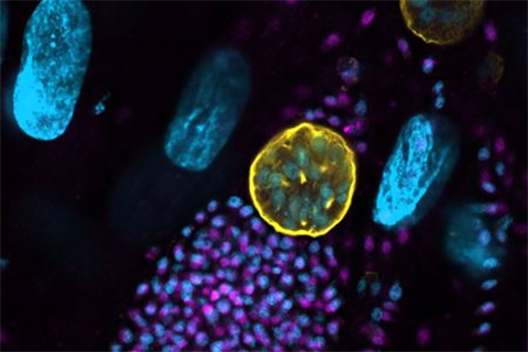
Toxoplasma gondii is a single-celled parasite that people catch from cat feces, unwashed produce or undercooked meat. The parasite has infected up to one-third of the world's population, and after causing mild illness, it persists by entering a dormant phase housed in cysts throughout the body, including the brain.
Toxoplasma cysts have been linked to behavior changes and neurological disorders like schizophrenia. They can also reactivate when the immune system is weakened, causing life-threatening organ damage. While drugs are available to put toxoplasmosis into remission, there is no way to clear the infection. A better understanding of how the parasite develops into cysts would help scientists find a cure.
Through years of collaborative work, IU School of Medicine professors Bill Sullivan, and Ronald C. Wek, have shown that Toxoplasma forms cysts by altering which proteins are made. Proteins govern the fate of cells and are encoded by mRNAs.
"But mRNAs can be present in cells without being made into protein," Sullivan said. "We've shown that Toxoplasma switches which mRNAs are made into protein when converting into cysts."
Lead author Vishakha Dey, a postdoctoral fellow at the IU School of Medicine and a member of the Sullivan lab, examined the so-called leader sequences of genes named BFD1 and BFD2, both of which are necessary for Toxoplasma to form cysts.
"mRNAs not only encode for protein, but they begin with a leader sequence that contains information on when that mRNA should be made into protein," Dey said.
All mRNAs have a structure called a cap at the beginning of their leader sequence. Ribosomes, which convert mRNA into protein, bind to the cap and scan the leader until it finds the right code to begin making the protein.
"What we found was that, during cyst formation, BFD2 is made into protein after ribosomes bind the cap and scan the leader, as expected," Dey said. "But BFD1 does not follow that convention. Its production does not rely on the mRNA cap like most other mRNAs."
The team further showed that BFD1 is made into protein only after BFD2 binds specific sites in the BFD1 mRNA leader sequence.
Sullivan said this is a phenomenon called cap-independent translation, which is more commonly seen in viruses.
"Finding it in a microbe that has cellular anatomy like our own was surprising," Sullivan said. "It speaks to how old this system of protein production is in cellular evolution. We're also excited because the players involved do not exist in human cells, which makes them good potential drug targets."
The Journal of Biological Chemistry featured the new study as an Editor's Pick, which represent a select group of the journal’s publications judged to be of exceptionally high quality and broad general interest to their readership.
This article is republished from the Indiana University School of Medicine Newsroom. Read the original here.
Enjoy reading ASBMB Today?
Become a member to receive the print edition four times a year and the digital edition monthly.
Learn moreGet the latest from ASBMB Today
Enter your email address, and we’ll send you a weekly email with recent articles, interviews and more.
Latest in Science
Science highlights or most popular articles

Glaucoma model links immune signaling to disease progression
Researchers at Duke University determine genetic variations that could increase the risk of developing glaucoma.

Uncovering the molecular roots of fatty liver disease
Physician–scientist Silvia Sookoian discusses her path from hepatitis C care to MASLD research, her use of multi-omics to study steatotic liver disease, and how lipid metabolism and genetics are reshaping understanding of MASH and liver health.

Mitochondria shape kidney cell function
Researchers at the University of Washington, Seattle present the first quantitative comparison of mitochondrial interactomes between two epithelial cell types in the kidney.

Long-chain polyunsaturated fatty acids linked to postoperative delirium risk
Researchers show that altered lipid metabolism may contribute to postoperative delirium, a condition linked to increased risk for long-term cognitive decline. The study explores potential disease mechanisms, which have yet to be understood.

Glycosylation patterns across antibody isotypes distinguish tuberculosis states
Researchers at Taipei Medical University present the first site-specific glycosylation analysis of immunoglobulins in elderly tuberculosis patients.

Blood glycome possibly predicts lifespan
Researchers at the University of Santiago de Compostela show that total serum N-glycome can predict mortality independent of traditional risk factors.