Sex and diet shape fat tissue lipid profiles in obesity

In obesity, adipose tissue expands and accumulates, driving chronic inflammation. Previous research showed that sex steroid hormones can influence adipose tissue distribution, accumulation and immune responses in men and women. Changes in lipid composition in the visceral or gonadal white adipose tissue, or GWAT, during obesity can drive immune cell accumulation and boost proinflammatory mediators. Prior studies revealed sex differences in GWAT lipid species in obese mice, but scientists still do not understand the exact role of sex hormones in lipid composition.
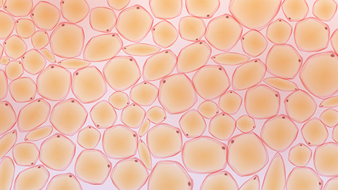
In a recent study in the Journal of Lipid Research, Mita Varghese and a team of researchers at the University of Michigan investigated the GWAT lipid profiles in obese mice and mice with their gonads surgically removed, or GX mice. In an untargeted lipidomics analysis where they comprehensively analyzed all lipids, they found sex differences in several lipid species, such as phospholipids and sphingolipids, which are important cell membrane components. Obese males had significantly more precursor fatty acids than females and GX mice. Targeted analysis revealed sex differences in polyunsaturated fatty acids, or PUFAs, with males showing a significantly higher omega-6 to omega-3 ratio. They also found diet-driven differences in oxylipins, inflammation-linked lipids, which were higher in both male and female obese mice than in lean mice.
This study suggests that sex hormone levels and diet equally induce inflammation and changes in lipid composition in obesity. Future studies include further confirming lipid profiles and understanding how sex differences arise in obesity.
Enjoy reading ASBMB Today?
Become a member to receive the print edition four times a year and the digital edition monthly.
Learn moreGet the latest from ASBMB Today
Enter your email address, and we’ll send you a weekly email with recent articles, interviews and more.
Latest in Science
Science highlights or most popular articles

Blood glycome possibly predicts lifespan
Researchers at the University of Santiago de Compostela show that total serum N-glycome can predict mortality independent of traditional risk factors.

Building a better model for drug delivery across the blood–brain barrier
Industry and academic scientists collaborated to develop a rat with humanized iron-transport receptors, enabling research into iron homeostasis and drugs that cross the brain’s barrier.

Fat synthesis enzyme crucial for milk fat and newborn growth
Researchers found that a deficiency of the fatty acid synthesis enzyme stearoyl-CoA desaturase-1 reduced mammary gland function during lactation and caused low birth weight in newborns that were fed milk from enzyme-deficient glands.

Flipping lipids and slime molds
A dull first job nearly pushed JBC associate editor Todd Graham out of science. Then a slime mold project changed his path. Now, he studies membrane biology and reflects on discovery, persistence and mentoring through uncertainty.

How smelling death alters worm behavior
Researchers have found that the roundworm C. elegans can smell death, and it changes how the worms behave, reproduce and age.

A chance encounter with the lab
Payton Stevens never planned to become a pancreatic cancer researcher. A temporary job set him on a path from rural Kentucky to leading research on Wnt signaling and metastasis, where he now pairs discovery with mentorship and science advocacy.