Helping mitochondria run smoothly may protect against Parkinson’s disease


In 1817, a British physician named James Parkinson published An Essay on the Shaking Palsy, describing for the first time cases of a neurodegenerative disorder now known as Parkinson’s disease. Today, Parkinson’s disease is the second most common neurodegenerative disease in the U.S. It affects about 1 million Americans and more than 10 million people worldwide.
The signature shaking in patients with the disease is the result of dying brain cells that control movement. To date, there are no treatments available that can stop or slow down the death of those cells.
We are researchers who study Parkinson’s disease. For over a decade, our lab has been investigating the role that mitochondria – the powerhouses that fuel cells – play in Parkinson’s.
Our research has identified a key protein that could lead to new treatments for Parkinson’s disease and other brain conditions.
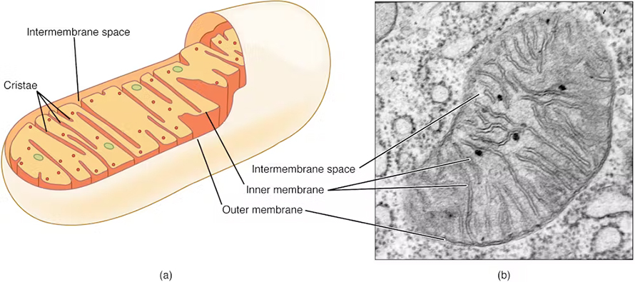
Mitochondrial dynamics and neurodegeneration
Unlike actual power plants, which are set in size and location, mitochondria are rather dynamic. They constantly shift in size, number and location, traveling between many different parts of the cells to meet different demands. These mitochondrial dynamics are vital to not only the function of mitochondria but also the health of cells overall.
A cell is like a factory. Multiple departments must seamlessly work together for smooth operations. Because many major processes interconnect, impaired mitochondrial dynamics could cause a domino effect across departments and vice versa. Collective malfunction in different parts of the cell eventually leads to cell death.
Emerging studies have linked imbalances in mitochondrial processes to different neurodegenerative diseases, including Parkinson’s disease. In many neurodegenerative disorders, certain disease-related factors, such as toxic proteins and environmental neurotoxins, disrupt the harmony of mitochondrial fusion and division.
Impaired mitochondrial dynamics also take down the cell’s cleaning and waste recycling processes, leading to a pileup of toxic proteins that form harmful aggregates inside the cell. In Parkinson’s, the presence of these toxic protein aggregates is a hallmark of the disease.
Targeting mitochondria to treat Parkinson’s
Our team hypothesized that restoring mitochondrial function by manipulating its own dynamics could protect against neuronal dysfunction and cell death.
In an effort to restore mitochondrial function in Parkinson’s, we targeted a key protein that controls mitochondrial dynamics called dynamin-related protein 1, or Drp1. Naturally abundant in cells, this protein travels to mitochondria when they divide into smaller sizes for higher mobility and quality control. However, too much Drp1 activity causes excessive division, leading to fragmented mitochondria with impaired function.
Using different lab models of Parkinson’s, including neuronal cell cultures and rat and mice models, we found that the presence of environmental toxins and toxic proteins linked to Parkinson’s cause mitochondria to become fragmented and dysfunctional. Their presence also coincided with the buildup of those same toxic proteins, worsening the health of neuronal cells until they eventually started dying.
We also observed behavior changes in rats that impaired their movements. By reducing the activity of Drp1, however, we were able to restore mitochondria to their normal activity and function. Their neurons were protected from disease and able to continue functioning.

In our 2024 study, we found an additional benefit of targeting Drp1.
We exposed neuronal cells to manganese, a heavy metal linked to neurodegeneration and an increased risk of parkinsonism. Surprisingly, we found that manganese was more harmful to the cell’s waste recycling system than to its mitochondria, causing buildup of toxic proteins before mitochondria became dysfunctional. Inhibiting Drp1, however, coaxed the waste recycling system back into action, cleaning up toxic proteins despite the presence of manganese.
Our findings indicate that inhibiting Drp1 from more than one pathway could protect cells from degeneration. Now, we’ve identified some FDA-approved compounds that target Drp1 and are testing them as potential treatments for Parkinson’s.
This article is republished from The Conversation under a Creative Commons license. Read the original article.
![]()
Enjoy reading ASBMB Today?
Become a member to receive the print edition four times a year and the digital edition monthly.
Learn moreGet the latest from ASBMB Today
Enter your email address, and we’ll send you a weekly email with recent articles, interviews and more.
Latest in Science
Science highlights or most popular articles

Uncovering the molecular roots of fatty liver disease
Physician–scientist Silvia Sookoian discusses her path from hepatitis C care to MASLD research, her use of multi-omics to study steatotic liver disease, and how lipid metabolism and genetics are reshaping understanding of MASH and liver health.

Mitochondria shape kidney cell function
Researchers at the University of Washington, Seattle present the first quantitative comparison of mitochondrial interactomes between two epithelial cell types in the kidney.

Long-chain polyunsaturated fatty acids linked to postoperative delirium risk
Researchers show that altered lipid metabolism may contribute to postoperative delirium, a condition linked to increased risk for long-term cognitive decline. The study explores potential disease mechanisms, which have yet to be understood.

Glycosylation patterns across antibody isotypes distinguish tuberculosis states
Researchers at Taipei Medical University present the first site-specific glycosylation analysis of immunoglobulins in elderly tuberculosis patients.

Blood glycome possibly predicts lifespan
Researchers at the University of Santiago de Compostela show that total serum N-glycome can predict mortality independent of traditional risk factors.

Building a better model for drug delivery across the blood–brain barrier
Industry and academic scientists collaborated to develop a rat with humanized iron-transport receptors, enabling research into iron homeostasis and drugs that cross the brain’s barrier.