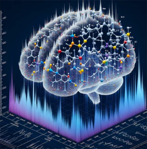
A proteomic hunt for phosphosites in the aging brain

Proteins are cellular workhorses, executing important tasks based on their specific functions. However, misfolded, mutated, inactive or overactive proteins are often the culprits behind disorders, including neurodegenerative diseases. Researchers need a greater understanding of how aging drives protein dysfunction to develop strategies to slow or even reverse these diseases.
Proteomics is broadly defined as the study of proteins — their structures, modifications, functions and abundance. The field has advanced significantly with the evolution of mass spectrometry, which allows scientists to identify and analyze proteins on a molecular level.
Uma Kanta Aryal, a research associate professor at Purdue University, identifies and analyzes changes in protein phosphorylation patterns and sites, also called phosphosites, which are significant contributors to Parkinson’s and Alzheimer’s diseases. He and his colleagues published their recent work in the journal Molecular & Cellular Proteomics.
Aryal’s interest in proteomics began in the early 2000s during his Ph.D. in Japan, where he used proteomic techniques to study enzymes from a white-rot fungus that degrade persistent aromatic compounds. He continued proteomics research through his postdocs and began his own proteomics program at Purdue in 2019 focusing on senescence, aging and their connection to neurodegenerative diseases.
“That’s where I switched,” Aryal said. “There is a lot of interest in neurodegenerative diseases, and there is a lot I can contribute to that area.”
Cells have mechanisms to activate and deactivate proteins, ensuring smooth cellular processes and preventing proteins from malfunctioning. Post-translational modifications, or PTMs, are critical in this regulation, though they often go underrecognized.
“Many people focus on proteins going up and down in abundance,” Aryal said. “But proteins are not only active when their expression goes up, it’s more related to their PTMs.”
Phosphorylation, a common PTM, is often implicated in disease. Protein hyperphosphorylation is responsible for aberrantly active proteins in several diseases. One example is tau, the protein that forms neurofibrillary tangles contributing to neuronal loss in Alzheimer’s and Parkinson’s diseases.
Aryal’s recent study looked at proteins in the brains of young, middle-aged and old mice using a multi-enzyme digestion approach for analysis by liquid chromatography–tandem mass spectrometry, or LC–MS/MS. This method breaks down proteins into smaller peptide pieces to identify hidden alterations: the PTMs. By examining these fragments, Aryal can detect and locate the subtle changes.
“There is a precise reason for each residue being modified,” he said. “That’s where I got excited — it’s not only the identification of those markers but characterizing them. Are they phosphorylated? If they are phosphorylated, where are they phosphorylated and what are the dynamics? How does this phosphorylation communicate with other PTMs?”
Aryal found increased abundance and activity of many kinases, enzymes that are responsible for phosphorylating other proteins, as well as changes in phosphorylation levels in proteins associated with neurodegeneration, including tau, Nefh and Dpysl2 in older mice.
Tau is widely studied due to its implication in Alzheimer’s and Parkinson’s diseases. Many phosphorylation sites of tau have already been mapped; however, Aryal located two additional tau sites in older mice.
“At one of these sites, the phosphorylation level goes up, while at the other the phosphorylation goes down in old mice,” Aryal said.
It’s almost like an internal struggle within the cell where one part of the tau protein seems to drive the development of diseases such as Alzheimer’s, and another part appears to counteract it. This dynamic suggests there’s still a lot we don’t know about tau and its role in neurodegeneration.
Aging is a major risk factor for all neurodegenerative diseases, and understanding the molecular mechanisms behind these conditions provides valuable insights for developing new therapies.
Aryal is now researching the changes in lipid profiles in the aging mouse brain and plans to use mice that have been genetically altered to have Alzheimer’s or Parkinson’s in future proteomic studies to better identify the disease states.
Enjoy reading ASBMB Today?
Become a member to receive the print edition four times a year and the digital edition monthly.
Learn moreGet the latest from ASBMB Today
Enter your email address, and we’ll send you a weekly email with recent articles, interviews and more.
Latest in Science
Science highlights or most popular articles

Mitochondria shape kidney cell function
Researchers at the University of Washington, Seattle present the first quantitative comparison of mitochondrial interactomes between two epithelial cell types in the kidney.

Long-chain polyunsaturated fatty acids linked to postoperative delirium risk
Researchers show that altered lipid metabolism may contribute to postoperative delirium, a condition linked to increased risk for long-term cognitive decline. The study explores potential disease mechanisms, which have yet to be understood.

Glycosylation patterns across antibody isotypes distinguish tuberculosis states
Researchers at Taipei Medical University present the first site-specific glycosylation analysis of immunoglobulins in elderly tuberculosis patients.

Blood glycome possibly predicts lifespan
Researchers at the University of Santiago de Compostela show that total serum N-glycome can predict mortality independent of traditional risk factors.

Building a better model for drug delivery across the blood–brain barrier
Industry and academic scientists collaborated to develop a rat with humanized iron-transport receptors, enabling research into iron homeostasis and drugs that cross the brain’s barrier.

Fat synthesis enzyme crucial for milk fat and newborn growth
Researchers found that a deficiency of the fatty acid synthesis enzyme stearoyl-CoA desaturase-1 reduced mammary gland function during lactation and caused low birth weight in newborns that were fed milk from enzyme-deficient glands.