Completing the cycle of genomic medicine
There are many challenges in diagnosing and treating rare diseases. A fundamental one is figuring out what is actually going wrong in a patient.

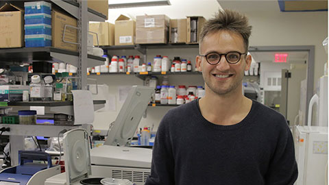
Wolfgang Pernice is a new assistant professor at Columbia University. He recently opened what he calls the Laboratory for Digital Biology, where his team is trying to address this hulking problem for Charcot‒Marie‒Tooth, or CMT, disease. He hopes his group’s solutions will apply to a host of other diseases too.
CMT is a disease of the nerves that serve the hands, arms, feet and legs. The nerves lose the ability to send strong signals to activate the muscles, which leads to the weakening of the muscles. This causes difficulty walking and carrying out everyday tasks. It can also be painful. There’s currently no cure and very limited treatments.
Pernice has CMT. He knows the disease and the problems in research from both the bench and bedside. “I know firsthand the techniques we’re using are not up to par,” he said of both the patient's struggle to get a diagnosis and the scientist's struggle to develop treatments.
Having the disease you’re trying to study adds a sense of urgency to your work. When Pernice was a Ph.D. student, he officially studied mitochondria and yeast biology, but on nights and weekends he worked with an additional mentor trying to figure out his own disease. Now he’s aiming to drive at the heart of the disease in his new lab.
Gaps in the cycle of genomic medicine
Medical research is often imagined as going from basic research, to preclinical, to clinical, tracing a line from lab benches all the way to patient bedsides. Pernice points out that it really starts in the other direction, from the bedside to the bench, making the process more of a loop or cycle than an arrow. It starts, he noted, with figuring out what is happening in an individual. In other words, before you can research a cure, you have to find out what, exactly and specifically, is wrong.
Ideally, you’d be able to diagnose people easily with some phenotype and genetic hallmark that makes things certain. Then, you’d have an easy thing to test all your potential medicines against, and you could come up with a cure.
“A lot of cases that we see in the clinic really don’t progress down the cycle: clinic, phenotyping, genetic diagnosis, benchwork, clinical trial, patient,” Pernice said.
On the diagnosis side, there’s what’s known as the “diagnostic odyssey,” in which patients are shuttled from doctor to doctor, specialist to specialist, trying to figure out a diagnosis, let alone a treatment. Many patients are misdiagnosed with conditions that have overlapping symptoms. Meanwhile, the lack of information about the specifics of the disease is a stumbling block on the research side.
From mutation to phenotype
“We can sequence people left and right,” Pernice said. But, unlike what happens in TV medical dramas, that doesn’t always give you an answer.
Imagine, for example, you’ve found five candidate genes from a group of patient sequences and you want to test them. What exactly are you testing for? If you delete a gene from a cell, what are you looking for to know if it’s the one causing the disease? If you add a potential treatment, what are you looking for to be fixed?
“The key thing you’re missing in order to get started is the phenotype,” Pernice said, and that is what he is working on. Researchers need a good and reliable readout in cell-based experiments for what is a disease and what is healthy.
Analyzing images of cells is one way you can begin to understand what’s happening.
Pernice spent time as a Ph.D. student doing image analysis by hand. He used a cursor to manually draw a circle on the microscope image and asked the program he was using to quantify the fluorescence inside it. “My whole Ph.D. was drawing circles,” he joked.
He knew that was not the way he wanted to go about analyzing images in the future. It was cumbersome and looked at only a very limited amount of data at a time.
His approach now is cell profiling, a high-content type of analysis inspired by Anne Carpenter at the Broad Institute, who does “cell painting”: imaging cells to collect as much information as you can.
Pernice is working on an automated, machine-learning way to find phenotypes in the images, once he’s collected as much visual data as possible from each cell.
He compares the approach to a human looking at a face. “We have evolved to look at someone’s face and get an idea of what they’re thinking. But you can’t necessarily tell you how you know,” he said, furrowing his eyebrow to demonstrate a facial expression people can generally read without having to stop and measure each other’s faces.
We don’t typically consciously quantify how high an eyebrow is raised, or when exactly a line around a mouth appears; we just usually get the emotion being conveyed from taking all the information in at once.
However, he explained, “We certainly haven’t evolved to see and observe and understand the microexpressions that the cells show. They’re amorphous, complicated.”
Pernice and his group have invested a good amount of time, several years already, in making algorithms that aim to read cells the way people read faces, looking for meaningful information.
They have collected cells from a group of patients with CMT, including himself. They’ve imaged the cells and fed the imaging data into the algorithm they’re developing. In return, it’s giving phenotypic information back. “That’s been really rewarding, and we’ve been able to identify some latent phenotypes in CMT subtypes,” he said.
“Subtypes” is key here. There are already two known subtypes of CMT: a demyelinating subtype, which shows a defect in nerve conduction velocity, and a subtype in which the axons themselves have a defect and the conduction velocity is normal but the amplitude of the signal is lower than it should be.
Besides this distinction, there are turning out to be many more subtypes. CMT, like many diseases, seems to be a constellation of diseases, all slightly different. There are more than a hundred genes that are associated with CMT, Pernice said, and there may turn out to be many more subtypes discovered by Pernice’s cell profiling data too.
Having so many subtypes means that a phenotype that is a hallmark of one subtype may not show up in another, or a gene that is causal in one may not be in another. This itself is critical information since once you begin testing hypotheses at the bench you want to know for certain what samples you are working with so you can test the right variables. A treatment for one subtype may not work for another, and being able to classify who belongs to what group can help identify who is likely to benefit from a treatment. Pernice hopes to use the platform to find phenotypes that are different from wild type for any patient that comes to the clinic.
Pernice said he is hopeful about the work of his team and that of other CMT researchers around the world who are making real progress. He said he sees a few possible treatments on the horizon. Together, scientists are beginning to close the gaps in the cycle from the patient, to the lab and back to the patient.
Enjoy reading ASBMB Today?
Become a member to receive the print edition four times a year and the digital edition monthly.
Learn moreGet the latest from ASBMB Today
Enter your email address, and we’ll send you a weekly email with recent articles, interviews and more.
Latest in Science
Science highlights or most popular articles

Key regulator of cholesterol protects against Alzheimer’s disease
A new study identifies oxysterol-binding protein-related protein 6 as a central controller of brain cholesterol balance, with protective effects against Alzheimer’s-related neurodegeneration.

From humble beginnings to unlocking lysosomal secrets
Monther Abu–Remaileh will receive the ASBMB’s 2026 Walter A. Shaw Young Investigator Award in Lipid Research at the ASBMB Annual Meeting, March 7-10 in Washington, D.C.

Chemistry meets biology to thwart parasites
Margaret Phillips will receive the Alice and C. C. Wang Award in Molecular Parasitology at the ASBMB Annual Meeting, March 7-10 in Washington, D.C.

ASBMB announces 2026 JBC/Tabor awardees
The seven awardees are first authors of outstanding papers published in 2025 in the Journal of Biological Chemistry.

Missing lipid shrinks heart and lowers exercise capacity
Researchers uncovered the essential role of PLAAT1 in maintaining heart cardiolipin, mitochondrial function and energy metabolism, linking this enzyme to exercise capacity and potential cardiovascular disease pathways.

Decoding how bacteria flip host’s molecular switches
Kim Orth will receive the Earl and Thressa Stadtman Distinguished Scientists Award at the ASBMB Annual Meeting, March 7–10, just outside of Washington, D.C.

