Chasing one of biology's biggest goals: Filming proteins in motion

Proteins are the heavy-lifters of biochemistry. These beefy molecules act as building blocks, receptors, processors, couriers and catalysts. “Proteins are the molecular machines that power all life on Earth,” explained Mark Sherwin, a physics professor at UC Santa Barbara. Naturally, scientists have devoted a lot of research to understanding and manipulating proteins.
A team led by researchers at UC Santa Barbara, including Sherwin, has made strides in addressing one of the grand challenges of modern science: recording proteins in motion in a lifelike environment. The authors discuss their technique in Angewandte Chemie, a journal of the German Chemical Society. The approach could revolutionize our understanding of how proteins do their jobs and guide the design of proteins for specific purposes.
A daunting challenge
Understanding a protein’s function requires more than merely a list of its parts. For these molecules, form begets function. Scientists have made enormous progress in the last 20 years deciphering proteins’ shapes based on the amino acid building blocks that form them.

However, even seeing a machine’s shape often isn’t enough to understand how it works. “Imagine you are an alien, and you see a picture of a sewing machine,” Sherwin said. “You would have a hard time figuring out what it does. But if you saw a movie, you would have a much better idea.”
Unfortunately, this is a tall order for proteins. Although they are relatively large molecules, proteins are still only a few nanometers in size, 100 times smaller than we can resolve even with the most powerful optical microscopes. And they exist in wet, squishy environments not conducive to cinematography.
“One of the biggest challenges in biology in general is to see proteins in action,” explained co-lead author Shiny Maity, a chemistry doctoral student. It is much easier for scientists to look at the structure of proteins when they’re frozen. To see them move requires a technique like stop-motion animation: start the action, freeze the protein, capture an image, repeat. This is often prohibitively difficult for both fast and slow movement. On top of that, flash freezing the protein can affect its structure.
“The goal that we have is to take out the freezing aspect completely and look at the motion of the protein in as close to a lifelike environment as we can get,” said co-lead author Brad Price, a physics graduate student.
An intricate technique
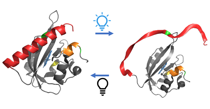
This paper demonstrates a new method to track the movement of proteins in a lifelike environment after their motion has been triggered by an external event (in this case, a visible light pulse). The authors call the technique TiGGER, for Time Resolved Gadolinium-Gadolinium Electron paramagnetic Resonance. It is elaborate, requiring quantum phenomena, skilled chemistry, specialty equipment and bioengineering.
TiGGER involves tagging two spots on the protein and tracking the distance between these labels as the protein unfolds and refolds. The star of the show is a charged gadolinium atom, or ion. Its electrons line up in such a way that the ion behaves like a little magnet. If you place it in a strong magnetic field, it will align with or against the external field and begin to wobble.
Scientists stick the gadolinium in a molecular cage to stabilize it and add some chemical scaffolding to link it with the protein. But those bits only link to one type of amino acid, cysteine. So the team had to change the amino acids they wanted to tag into cysteines without affecting the protein’s overall function. It was a task made even trickier by a cysteine in the center of the protein that is critical to its function.
“The spin label is chosen very strategically,” Maity said. “It is big enough not to enter the core of the protein, where the functional cysteine is located. But it’s also not too big that it disrupts the protein’s natural shape.”
The wobbling, or “precession,” of the gadolinium ion is influenced by the proximity of the other tag, which has its own wobbling gadolinium ion generating its own little magnetic field. This precession changes based on how close the two tags are to each other. Measure this wobble, and you can derive the distance.
This is precisely what the authors did using a laser with light at energies slightly higher than those in a microwave oven. When the frequency of these sub-Terahertz waves and the ion’s precession match up, the waves are absorbed. The scientists then measured this absorption to detect small changes in the gadolinium’s precession. If the amount of absorption changes with time, that means the tags are moving.
Add some more mathematics, and the authors could even tell you how far apart the tags are from one another. “We know that we can get distance as a function of time, but it will take more development,” Price said.
An illuminating protein
The authors selected a popular and versatile protein to develop TiGGER. Their model belongs to the light, oxygen or voltage (LOV) sensitive family of proteins, specifically a light-activated protein called AsLOV2. “LOV proteins control processes ranging from circadian rhythms in bacteria, plants and fungi to phototropism in plants and microorganisms,” said co-author Max Wilson, an assistant professor in the Department of Molecular, Cellular and Developmental Biology. “In summary, they are intimately connected to light sensing.”
This property makes AsLOV2 popular with scientists and engineers, and simple to manipulate. “It’s interesting and a perfect test case,” Price said, “a best-of-both-worlds situation.”
LOV proteins enable scientists to use light as a “remote control” for a whole host of molecular processes in cells. “We use it to control stem cell differentiation, antibody binding, stiffening and relaxation of extracellular matrix proteins, and activation of cellular signaling pathways,” Wilson said.
Assistant professor Arnab Mukherjee, of the chemical engineering department, uses LOV proteins to track biochemical processes in living cells using fluorescence, much like a highlighter under a black light. “Unlike conventional fluorescent proteins, LOV proteins operate by a distinct mechanism that makes their ‘glow’ visible even in oxygen-free conditions,” he explained. This offers a tool to study microbes living in anaerobic environments, like the human gut.
But it’s tricky to engineer these proteins to do what researchers want. This is where TiGGER comes in handy. If scientists like Wilson and Mukherjee can see the proteins in motion, they could be more deliberate in their design processes.
An eye toward the future
Senior authors Sherwin and Songi Han, a professor of chemistry, first began their quest to film proteins in 2006, however it’s still early days for TiGGER. Right now, the technique can produce a one-dimensional trajectory of a protein’s movement between two points. But its true power comes from repeating the technique at several different sites. This enables scientists to piece together the motion of the protein as a whole. They can then map this movement onto a model of the protein to create a movie in a similar manner to the CGI animation that brings our favorite cartoon characters to life.
The authors are focused on optimizing the technique before they invest the time applying it to other sites on AsLOV2. They are working to increase the signal-to-noise ratio and bump up the sampling speed of their instruments. The team also hopes to slow down the random motion of the proteins as they’re suspended in solution, which should enable them to capture sharper footage than they can now.
In the meantime, Price and Maity are using TiGGER to answer some basic questions about AsLOV2. For instance, why does the protein unfold over 1,000 times faster than it refolds? And how do mutations that are known to affect refolding affect unfolding? They’re also investigating how hotter conditions affect the protein’s function. The results could shed light on how oats — the source of AsLOV2 — will respond to climate change.
Eventually, TiGGER can be translated to all sorts of other proteins, as long as the scientists can modify the sites of interest into cysteine amino acids without affecting the protein’s function. “Biophysicists have been striving to ‘film’ proteins in motion to gain an in-depth understanding of their biological functions,” Maity said. “TiGGER has the potential to make this dream come true.”
This article was first published by the University of California, Santa Barbara. Read the original.
Enjoy reading ASBMB Today?
Become a member to receive the print edition four times a year and the digital edition monthly.
Learn moreGet the latest from ASBMB Today
Enter your email address, and we’ll send you a weekly email with recent articles, interviews and more.
Latest in Science
Science highlights or most popular articles

Glaucoma model links immune signaling to disease progression
Researchers at Duke University determine genetic variations that could increase the risk of developing glaucoma.

Uncovering the molecular roots of fatty liver disease
Physician–scientist Silvia Sookoian discusses her path from hepatitis C care to MASLD research, her use of multi-omics to study steatotic liver disease, and how lipid metabolism and genetics are reshaping understanding of MASH and liver health.

Mitochondria shape kidney cell function
Researchers at the University of Washington, Seattle present the first quantitative comparison of mitochondrial interactomes between two epithelial cell types in the kidney.

Long-chain polyunsaturated fatty acids linked to postoperative delirium risk
Researchers show that altered lipid metabolism may contribute to postoperative delirium, a condition linked to increased risk for long-term cognitive decline. The study explores potential disease mechanisms, which have yet to be understood.

Glycosylation patterns across antibody isotypes distinguish tuberculosis states
Researchers at Taipei Medical University present the first site-specific glycosylation analysis of immunoglobulins in elderly tuberculosis patients.

Blood glycome possibly predicts lifespan
Researchers at the University of Santiago de Compostela show that total serum N-glycome can predict mortality independent of traditional risk factors.