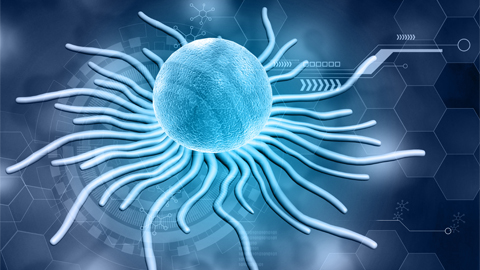
Peering into ocular waste recycling

A recent study in the Journal of Biological Chemistry revealed the key to a protein that commonly causes blindness. The biological process involves a protein that is essential for transporting toxic compounds out of the eye, similar to a garbage recycling service. The challenge is that, like food and the waste it generates, these compounds are essential for the eye to function properly — until they build up and cause blindness.

The scientists behind the study research a protein transporter, called ABCA4, that lines the edges of specialized photoreceptor cells in the retina and is normally poised to remove toxic, fatty retinal byproducts called N-Ret-PE. Retinal is a derivative of vitamin A, which is found in foods such as leafy green vegetables.
“Retinal is critical for vision,” said Robert Molday, a professor of biochemistry and molecular biology at the University of British Columbia who oversaw the work. “But, it's also potentially very toxic because it has a very reactive element. So, cells have to be able to balance between using retinal for sustained vision as well as managing its toxicity .”
Mutations in ABCA4 can cause N-Ret-PE buildup, which leads to vision loss in diseases such as Stargardt disease. Stargardt disease is the most common inherited form of macular degeneration and affects approximately 30,000 people nationwide. There is currently no therapy or cure for the disease.

The researchers were interested in finding out how the ABCA4 transporter malfunctions to cause vision loss. They found that a portion of the protein that interacts with N-Ret-PE, known as the binding pocket, is inert in some patients with Stargardt disease. Therefore, the toxic compounds slip out of the ABCA4 transporter and cannot be removed from the retina.
Next, by changing the makeup of ABCA4, the researchers showed they could mimic the effect of the Stargardt mutations.
“We were able to elucidate the mechanism of binding, which paves the way for treatments for Stargardt disease,” Tongzhou Xu, a postdoctoral fellow at UBC and lead author of the study, said.
The team is optimistic that one day there will be a targeted therapeutic for patients with Stargardt disease that may use gene therapy and specialized particles for delivery to the eye. Gene therapy approaches have already been successfully used to correct mutations in a similar transporter, which causes cystic fibrosis.
“We are now applying two types of technologies to alter ABCA4,” Molday said. “One which was developed to specifically correct the DNA with gene-editing approaches. We are coupling that with lipid nanoparticles, which have been used in the COVID-19 vaccine to encapsulate mRNA. So, by combining these two technologies, we envision being able to potentially correct the defects in individuals with Stargardt's disease that have specific point mutations.”
Enjoy reading ASBMB Today?
Become a member to receive the print edition monthly and the digital edition weekly.
Learn moreGet the latest from ASBMB Today
Enter your email address, and we’ll send you a weekly email with recent articles, interviews and more.
Latest in Science
Science highlights or most popular articles
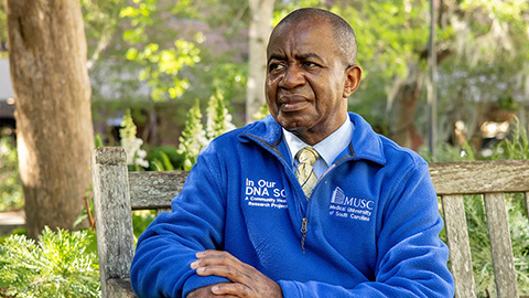
Genetics studies have a diversity problem that researchers struggle to fix
Researchers in South Carolina are trying to build a DNA database to better understand how genetics affects health risks. But they’re struggling to recruit enough Black participants.
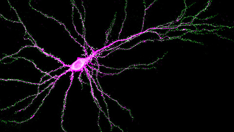
Scientists identify new function of learning and memory gene common to all mammalian brain cells
Findings in mice may steer search for therapies to treat brain developmental disorders in children with SYNGAP1 gene mutations.
From the journals: JBC
Biased agonism of an immune receptor. A profile of missense mutations. Cartilage affects tissue aging. Read about these recent papers.
Cows offer clues to treat human infertility
Decoding the bovine reproductive cycle may help increase the success of human IVF treatments.
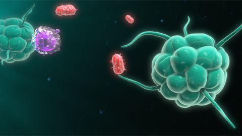
Immune cells can adapt to invading pathogens
A team of bioengineers studies how T cells decide whether to fight now or prepare for the next battle.
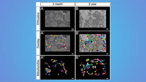
Hinton lab maps structure of mitochondria at different life stages
An international team determines the differences in the 3D morphology of mitochondria and cristae, their inner membrane folds, in brown adipose tissue.