Progress in identifying lipid domains (rafts) in living cells

Under which conditions lipid chemical heterogeneity results in the formation of coexisting lipid domains with distinct lipid compositions and properties in living cells has been a subject of intense research for decades.
In model membrane formed from lipid mixtures, spontaneous formation of tightly packed sphingolipid- and cholesterol-rich lipid domains (in the liquid-ordered state) that segregate from loosely packed domains richer in unsaturated phospholipids (in the liquid-disordered state) are detected and characterized easily.

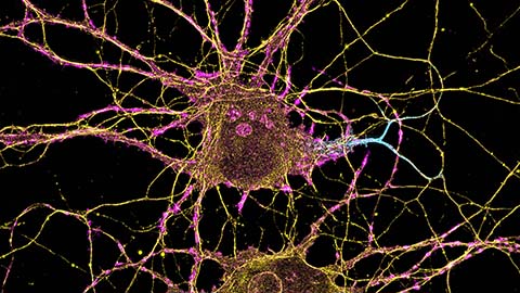
However, analogous domains in cells are very small under most conditions — at or beyond the limit of detection for most techniques. This has led to much controversy as well as much work aiming to develop new methods to identify and characterize tiny nanodomains.
Very recent progress in living cells has been encouraging on several fronts. Studies using novel fluorescently labeled lipids with affinities for liquid-ordered domains similar to those of unlabeled lipids have revealed that specific association of raft-loving lipids with raft-localizing proteins occurs in living cells (1,2). Single-particle-tracking measurements show that these interactions are lost in living cells when even minor changes in lipid or protein structure are made if these changes abolish raft-associating physical properties.
In other studies, super-resolution microscopy in B cells has found co-localization of raft markers with, and exclusion of nonraft markers from, the vicinity of clustered B-cell receptors on a size scale similar to that of the clusters (50 nanometers to 100 nanometers). This is indicative of the formation of ordered domains around the B-cell receptors. An analogous formation of nanodomains was detected around clustered cholera toxin, a molecule long known to induce the formation of ordered domains in vitro and in cells (3).
These studies extend previous work from other labs that reported lipid-domain-based molecular interactions in these systems. This is indicative of a robust underlying phenomenon.
Advances leading to an increased ability to visualize domains and manipulate their structure promise further progress. An even higher-resolution, super-resolution microscopy approach has been developed, which may allow visualization of domains that otherwise would elude direct visualization (4).
Finally, our own lab has devised a method efficiently to replace virtually the entire complement of plasma membrane outer leaflet lipids in living cells with exogenous lipids. This may allow fine-tuned control of domain formation and properties (5).
REFERENCES
1. Komura, N. et al. Nat. Chem. Biol. 12, 402 – 410 (2016).2. Kinoshita, M. et al. J. Cell. Biol. 216, 1183 – 1204 (2017).
3. Stone, M.B. et al. eLife 6, e19891 (2017).
4. Balzarotti, F. et al. Science 355, 606 – 612 (2017).
5. Li, G. et al. Proc. Natl. Acad. Sci. USA 113, 14025 – 14030 (2016).
Enjoy reading ASBMB Today?
Become a member to receive the print edition four times a year and the digital edition monthly.
Learn moreGet the latest from ASBMB Today
Enter your email address, and we’ll send you a weekly email with recent articles, interviews and more.
Latest in Science
Science highlights or most popular articles

How smelling death alters worm behavior
Researchers have found that the roundworm C. elegans can smell death, and it changes how the worms behave, reproduce and age.

A chance encounter with the lab
Payton Stevens never planned to become a pancreatic cancer researcher. A temporary job set him on a path from rural Kentucky to leading research on Wnt signaling and metastasis, where he now pairs discovery with mentorship and science advocacy.

Light-activated small molecule could transform eye infection treatment
Contact lenses raise the risk of infectious keratitis, a leading cause of blindness worldwide. A biotech company is commercializing a light-activated therapy using a ROS-generating molecule to rapidly kill microbes in the cornea to preserve vision.
The molecular orchestra of memory
Calcium, calmodulin and calcium/calmodulin-dependent kinase II form a molecular axis that turns fleeting neural activity into lasting memories. New research shows how memories are stabilized, and possibly even protected or repaired.

Differences in pili structure modulate bacterial behavior
Researchers demonstrate how small changes in the structure of hair-like protein appendages can affect the behavior of Acinetobacter bacteria.

Cholesterol regulatory genes predict liver transplant outcomes
Researchers identify a link between cholesterol-regulating genes and liver transplant success, which could improve donor screening and patient outcomes.