David Goodsell:
The master of mol art
.jpg?lang=en-US&width=300&height=300&ext=.jpg)
Visualization of cells and macromolecules is indispensable for understanding their function. Whether we are looking at the cellular compartments or trying to understand a certain biological process at the molecular level, the power of a visual image is enormous. Direct visual methods such as light and electron microscopy reveal a coarse view of the cellular structure. X-ray crystallography and NMR allow us to get atomic details of the individual molecules. Biochemistry and biophysical methods provide quantitative information about isolated systems. Despite having all these tools and information, we still miss a complete view of a cell on the nanometer scale (1).
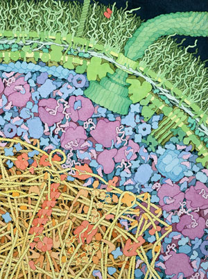 Fig 1. Cross section of an Escherichia coli. A large flagellar motor (in green) crosses the cell wall, turning the flagellum. The cytoplasmic area is colored blue and purple. The large magenta molecules are ribosomes, and the L-shaped maroon molecules are tRNA. The nucleoid region is shown in yellow and orange. Courtesy of D.S. Goodsell.
David Goodsell of The Scripps Research Institute, an artist by nature and scientist by training, bridges that knowledge gap and takes us into the invisible world of a cell. In his drawings, he beautifully combines scientific information from many different methods with his artistic vision.
Nature and nurture
Goodsell started painting early in his childhood, taught by his grandfather, who was an accomplished artist. In college, Goodsell majored in both chemistry and biology but not in art. His graduate school years (with Richard Dickerson at the University of California, Los Angeles) coincided with the increased use of computers in structural biology. While writing molecular graphics programs to visualize crystal structures, Goodsell became increasingly interested in scientific illustration. His professional interest in molecules gradually intertwined with the artistic desire to paint them. He credits his postdoctoral adviser, Arthur Olson (at Scripps), for creating an atmosphere where Goodsell could develop his skills as both a scientist and an artist.
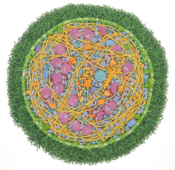 Fig. 2. Mycoplasma mycoides. DNA is shown in orange, cytoplasmic proteins in blue and pink, ribosomes are purple, and lipoglycan layer is green. Courtesy of D.S. Goodsell.
Artistic style
Goodsell uses watercolor to reveal a dazzling view of the molecular world inside living cells. His drawings usually are printed at a consistent 1,000,000x magnification, which enlarges the objects to visible size. He uses a cross section metaphor, which allows viewing of many molecules in the same image plane (2). This simplicity is somewhat reminiscent of the post-impressionist style of Henri Rousseau. Goodsell draws molecules with flat colors and outlines to emphasize their relative sizes. These artistic choices simplify the view of a bustling intracellular world.
E. coli (Fig. 1) is one of Goodsell's favorite subjects. Knowing a stunning amount of information about this bacterium allows the artist "to show everything that is needed to make a living object." Goodsell likes to use simple color schemes to highlight the function and location of the molecules. Whether he draws a bacterium or a human cell, it is easy to identify compartments by looking at the colors. DNA and nuclear proteins are drawn yellow and orange, ribosomes are shown in magenta, cytoplasmic proteins are shown in blue, and the membranes are colored green. The overall size and shape of the macromolecules are based on atomic coordinates. The relative amount of molecules is derived from biochemical data, and their locations are from electron micrographs.
To appreciate the amount of effort spent on a drawing, one has only to zoom into a recent painting of a Mycoplasma mycoides (Fig. 2). A close look at the elaborate lipoglycan layer alone surely will leave you amazed.
 Fig. 3. Neuromuscular synapse. The acetylcholine-laden vesicles are carrying and releasing the neurotransmitter into the synaptic cleft. A few of the acetylcholine molecules bind to receptors on the muscle cell. The cleft itself is packed with many elongated proteins including laminin, collagen, perlecan and flower-like acetylcholinesterase molecules serving to render inactive the neurotransmitter. Courtesy of D.S. Goodsell.
Paintings can tell great stories. Goodsell often uses his illustrations to describe a biological process vividly. His painting of the neuromuscular synapse, for example, shows the molecular action of the entire synaptic cycle (Fig. 3). An expert in vesicular trafficking, structural biologist Frederick Hughson of Princeton University concludes, "David's work is amazing!"
Science outreach
If you visit the Protein Data Bank website, you will not miss the "Molecule of the Month" column posted on the home page. Every month for more than a decade, David Goodsell has been contributing an illustration and a concise description of a featured biological macromolecule, such as clathrin (Fig. 4). Most of the structural images are created using a computer program that Goodsell developed as a postdoctoral fellow. With more than 60,000 structures currently deposited in the PDB, Goodsell has plenty of work to do.
Usually, figures appearing in original scientific publications must strictly represent the data, and thus very little image manipulation is allowed. However, artistic freedom is essential for a comprehensible and memorable illustration used for educational purposes (3), and several of Goodsell's images can be found in classic textbooks, such as "Molecular Biology of the Cell." In addition, Goodsell himself has written and illustrated a number of books, including "The Machinery of Life," "Our Molecular Nature: The Body's Motors, Machines and Messages" and "Bionanotechnology: Lessons from Nature."
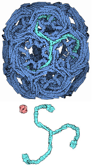 Fig. 4. Clathrin (April 2007 Molecule of the Month). A clathrin cage composed of 36 symmetrical three-legged components termed triskelions (single subunit is highlighted in green). The assembly shown here represents the second smallest possible cage structure. A hemoglobin molecule (red) is included for comparison. Courtesy of D.S. Goodsell.
Illustrations by Goodsell are making a great impact on science education. Microbiologist David Rudner at Harvard Medical School uses Goodsell's images in his molecular biology of bacteria course and finds them "incredibly instructive – both mnemonic and predictive… They stimulate general discussion and foster hypotheses." Alice Ting, a professor at MIT, also uses Goodsell pictures for teaching and in her seminars to illustrate how crowded and complex the interior of the cell is. "Other illustrations don't convey this nearly as well as the Goodsell drawings," she notes.
Goodsell recently has contributed a series of articles to the journal Biochemistry and Molecular Biology Education. In each paper, he describes in great detail the scientific background behind the illustrations in the second edition of "The Machinery of Life." BAMBED editors Judith and Donald Voet agree that Goodsell's work provides "enormous aid to the teaching and learning of biochemistry and molecular biology" (4).
As Goodsell says, his artwork is meant to give any reader "a pictorial overview of the molecules that orchestrate the process of life." For the scientist, he hopes that his work "will continue to provide a touchstone for intuition" (5).
References
1. Nogales, E. (2010) My dream of a fantastic voyage to see the inner workings of a cell. Mol. Biol. Cell.21, 3815.
2. Goodsell, D.S. (2005) Visual methods from atoms to cells. Structure 13, 347 – 354.
3. Goodsell, D.S., and Johnson, G.T. (2007) Filling in the gaps: artistic license in education and outreach. PLoS Biol.5, e308.
4. Voet, J.G., and Voet, D. (2009) Communication through illustration: The work of David Goodsell. Biochem. Mol. Biol. Educ. 37, 203.
5. Goodsell, D.S. (2009) The Machinery of Life. 2nd ed. Springer.
Enjoy reading ASBMB Today?
Become a member to receive the print edition monthly and the digital edition weekly.
Learn moreGet the latest from ASBMB Today
Enter your email address, and we’ll send you a weekly email with recent articles, interviews and more.
Latest in People
People highlights or most popular articles

2024 voter guide
Learn about the candidates running for ASBMB Council, Nominating Committee, Publications Committee and treasurer.
Charles O. Rock (1949 – 2023)
Colleagues and trainees remember a world expert in membrane lipid homeostasis.

Honors for Clemons, Hatzios and Wiemer
Awards, honors, milestones and more. Find out what's happening in the lives of ASBMB members.
Touching the future from the bench
Scholar, scientist, teacher and mentor Odutayo Odunuga discusses the important roles of the institutional PI, his journey and his research.

In memoriam: Darwin Prockop
He held leadership positions at multiple institutions and was known for his contributions to adult stem cell biology and cellular biology.

A look into medical writing
Our careers columnist spoke with Ashlea A. Morgan at Chameleon Communications International to get a sense of one type of work a medical writer can do.